
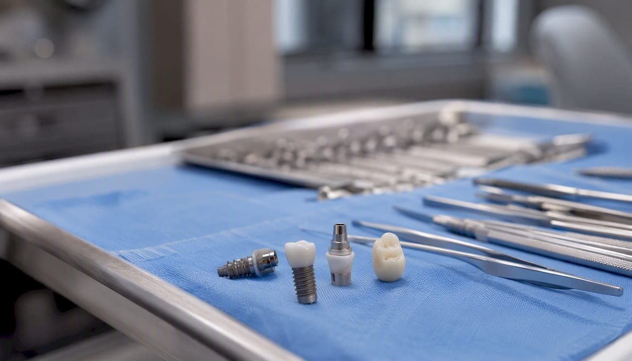
Yes, you can have an MRI if you have dental implants. Most modern implants are made from titanium or zirconia, neither of which is ferromagnetic, so the MRI magnet won't pull them out of your jaw. The real issue isn't patient safety — it's image quality. Implants can distort MRI images in the area around them, which matters most for head and neck scans.
Before your scan, do three things:
- Disclose your implants during the pre-scan screening call or at the intake desk. Tell the MRI team exactly what you have, including crowns, bridges, or any other dental restorations.
- Bring your implant card or documentation if you have it. The manufacturer's instructions for use (IFU) tell the radiology team whether your specific implant is classified MR-Safe, MR-Conditional, or MR-Unsafe.
- Remove any removable prostheses — dentures, partials, retainers, or removable orthodontic appliances — before entering the MRI room.
Standard medical protocol requires disclosure of all dental work before MRI, and many major implant systems are already classified MR-Conditional, meaning they can be scanned safely under specified conditions.
Key Takeaways
Most patients with dental implants can safely undergo MRI because titanium and zirconia are non-ferromagnetic; the primary concern is image artifact near the implant, not patient harm.
| Point | Details |
|---|---|
| MRI is safe for most implants | Titanium and zirconia implants are non-ferromagnetic; displacement and heating risks are minimal for well-integrated implants. |
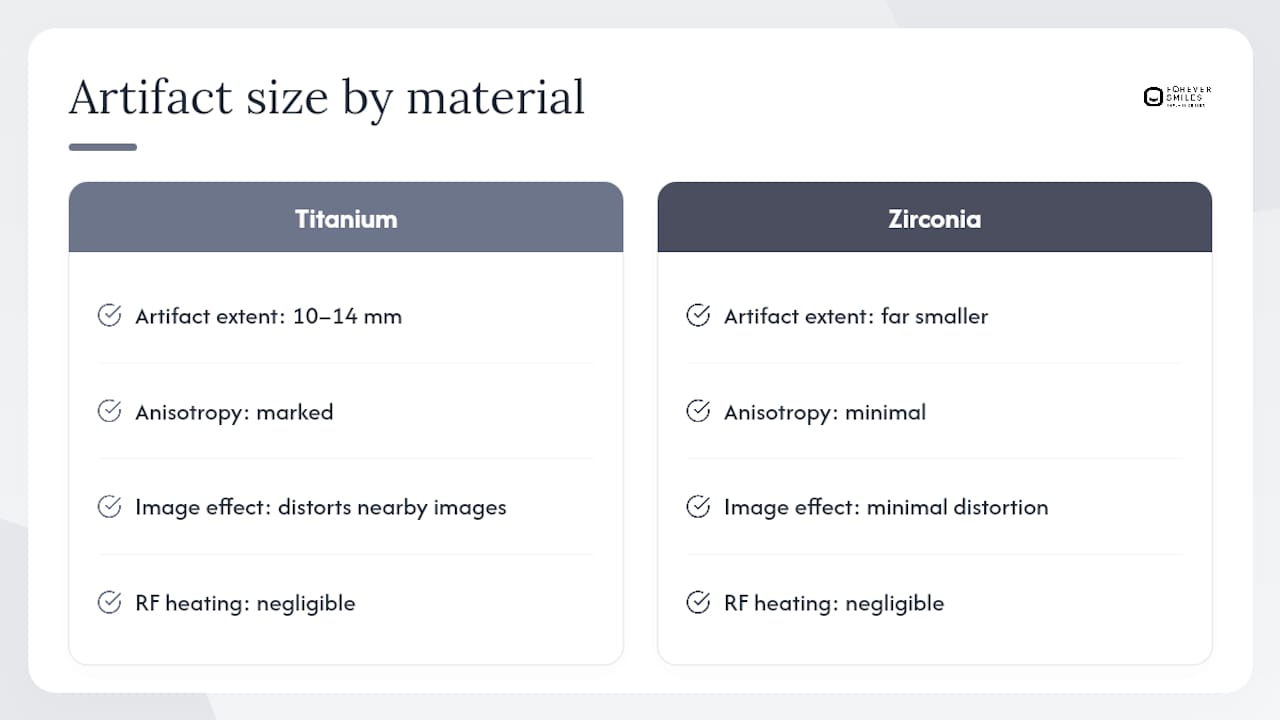
| Artifact, not safety, is the real issue | Titanium artifacts can extend 10–14 mm from the implant; zirconia produces markedly smaller distortion. |
| Always disclose and bring documentation | Tell the MRI team about all implants and restorations; bring your implant card or manufacturer IFU to confirm MR-Conditional parameters. |
| Red flags require coordination | Recent surgery, a loose implant, or unknown alloy composition warrants contact with your implant surgeon before scanning. |
| Radiology teams can reduce artifacts | Sequence selection, 1.5T vs 3T field strength, and metal artifact reduction techniques can improve image quality near implants. |
Table of Contents
- What are dental implants made of, and why does it matter for MRI?
- How does MRI actually interact with your implants?
- When do implants reduce MRI image quality, and what can the radiology team do?
- Pre-MRI checklist: what to tell the MRI team before your scan
- When should an MRI be delayed or modified because of dental work?
- A note from the implant team at Forever Smiles Implant Center
- What do MR-Safe, MR-Conditional, and MR-Unsafe mean for your implant?
- A message from Forever Smiles Implant Center
- Sources
What are dental implants made of, and why does it matter for MRI?
The material inside your mouth determines both how safe the scan is and how much it distorts the image.
Implant bodies are almost universally made from commercially pure titanium, titanium alloys (Ti-6Al-4V), or titanium-zirconium (Ti-Zr). These metals are paramagnetic, not ferromagnetic. That distinction is critical: ferromagnetic metals (like certain stainless steels) are strongly attracted to magnetic fields and can move or heat dangerously. Paramagnetic metals like titanium have a negligible magnetic response, which is why well-integrated titanium implants don't get ripped from the bone during a scan.
Zirconia implants are ceramic and essentially non-magnetic. Comparative studies show zirconia produces markedly smaller MRI artifacts than titanium or Ti-Zr alloys, making it the better option when MRI compatibility is a priority.
Restorations on top of implants are a different story. Porcelain-fused-to-metal (PFM) crowns contain metal substructures, and some older restorations use stainless steel or cobalt-chromium alloys that can cause more significant image distortion. Gold alloys are generally non-ferromagnetic but still produce some artifact. All-ceramic or zirconia crowns create the least interference.
Stainless steel orthodontic brackets and long-span metal-framed prostheses sit at the higher end of the concern spectrum. They aren't necessarily dangerous, but they can make head and neck MRI images difficult to read.
How does MRI actually interact with your implants?
Three separate things happen when you enter an MRI scanner with metal in your mouth. Understanding each one helps clarify what's actually at risk.
Displacement refers to the magnet physically moving or rotating the implant. For a well-osseointegrated titanium implant, this is rarely a concern. Clinical evidence confirms that displacement is rarely observed for well-fixed implants and that RF heating is generally negligible in standard clinical MRI. The bone that has grown around the implant holds it firmly in place.
RF heating occurs when radiofrequency energy from the scanner is absorbed by metal, potentially raising its temperature. For small titanium implants in typical 1.5T or 3T clinical scanners, the temperature rise is minimal and not considered clinically significant.
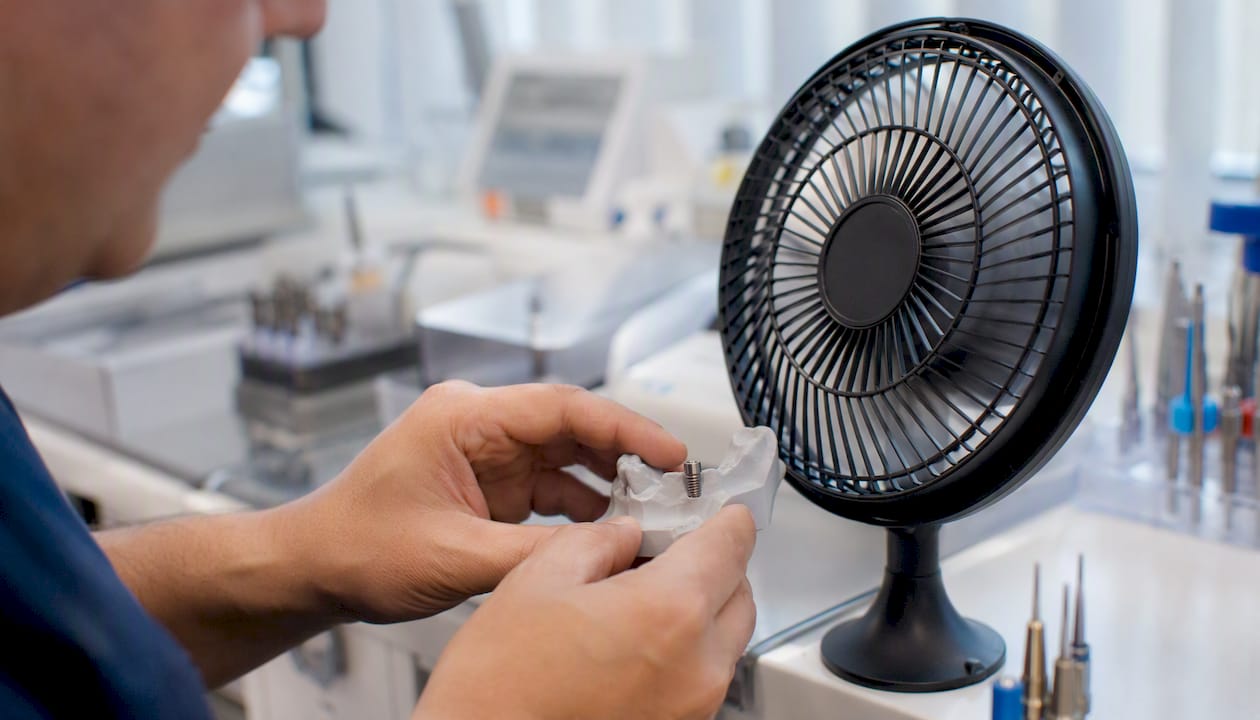
Susceptibility artifacts are the real problem. Metal disrupts the local magnetic field, causing signal voids and geometric distortions in the image. Titanium artifacts can extend 10–14 mm from the implant surface depending on the MRI sequence and the implant's orientation relative to the main magnetic field. The artifact geometry is often anisotropic, meaning it's larger in one direction than another. Zirconia produces far less distortion. The clinical consensus has shifted accordingly: patient safety risk from dental implants in MRI is minimal; the real problem is diagnostic quality when the area of interest is close to the implant.
When do implants reduce MRI image quality, and what can the radiology team do?
The primary clinical issue is artifact, not harm. Whether that artifact matters depends entirely on what the scan is trying to see.
Scans most likely to be affected by dental implant artifacts include:
- Brain MRI, particularly for structures near the skull base
- Paranasal sinus imaging
- Maxillofacial and oral soft tissue studies
- Temporomandibular joint (TMJ) evaluation
- Neck and cervical spine imaging near the jaw
A systematic review found that orthodontic appliances, especially stainless-steel brackets, commonly produce artifacts affecting head and neck MRI diagnosis. For dental implants and prosthetic restorations, the evidence is more variable, but material, location, and MRI parameters all determine severity.
Radiology teams have several tools to reduce artifact impact:
- Sequence selection: Spin-echo sequences produce smaller artifacts than gradient-echo sequences for metallic implants.
- Lower field strength: A 1.5T scanner generally produces less artifact than a 3T scanner for the same implant. When diagnostic quality near the implant is critical, 1.5T is often the better choice.
- Metal artifact reduction techniques (MARS): Specialized pulse sequences and reconstruction algorithms can suppress susceptibility artifacts significantly.
- Adjusted field of view and coil placement: Positioning the imaging coil away from the implant site and adjusting the FOV can reduce the artifact's impact on the region of interest.
Clinical reviews confirm that sequence choice, field strength, and metal artifact reduction techniques are the most practical mitigations available. Knowing the implant material helps the imaging team predict artifact size and choose the right approach.
Pre-MRI checklist: what to tell the MRI team before your scan
Walk into your MRI appointment prepared. The radiology team can only protect you and optimize your scan if they know what's in your mouth.
- Disclose all implants and restorations. Tell the MRI tech about every implant, crown, bridge, and any metal-containing restoration, not just the implants themselves.
- Bring your implant card or IFU. The manufacturer's documentation lists the MR safety classification and any scanning conditions that apply. If you don't have it, contact your implant surgeon's office before the appointment.
- Remove all removable dental appliances. Dentures, partial dentures, retainers, and removable orthodontic devices should come out before you enter the scanner room. Yale Medicine's MRI safety guidance specifically notes that removable metal appliances should be taken out when possible.
- Report any recent implant surgery. If your implant was placed within the past few weeks, tell the team. Healing implants warrant extra caution.
- Report any looseness. A loose implant or prosthesis changes the risk calculation.
- Disclose all other implanted devices. Pacemakers, cochlear implants, neurostimulators, and other devices may be MR-Unsafe and take priority in the screening process.
Pro Tip: When you call to schedule your MRI, use this script: "I have dental implants and fixed restorations. I have my implant manufacturer documentation available. Can you confirm what information your team needs before the scan?" That one sentence gets the right conversation started before you arrive.
When should an MRI be delayed or modified because of dental work?
Most patients with dental implants can proceed with MRI without delay. A few specific situations require a pause and a call to your dentist or implant surgeon.
Red flags that warrant dental or radiology coordination before scanning:
- Recently placed implants. During the healing period, osseointegration is incomplete. The implant is not yet fully anchored, and the risk profile differs from a mature, fully integrated implant. Contact your implant surgeon for guidance on timing.
- A loose implant or prosthesis. Any mobility in the implant or the restoration attached to it needs to be evaluated before MRI.
- Unknown metal composition. Long-span metal-framed bridges or older restorations placed decades ago may contain alloys whose magnetic properties are unknown. Without documentation, the radiology team can't confirm safety.
- Extensive stainless-steel prostheses or orthodontic appliances. These can produce significant artifacts and, in some configurations, may warrant alternative imaging.
- Other MR-Unsafe implanted devices. If you have a pacemaker, certain cochlear implants, or other devices classified MR-Unsafe, those take precedence over dental concerns entirely.
If any of these apply, contact your implant surgeon or dentist and request the manufacturer IFU for your specific implant system. For head and neck studies where artifact would make the scan unreadable, the radiologist may recommend CT or cone-beam CT (CBCT) as an alternative. Mayo Clinic's MRI safety guidance outlines the standard pre-scan screening process and the importance of disclosing all implanted devices.
A note from the implant team at Forever Smiles Implant Center
Dr. Brian Young is a residency-trained implant surgeon with more than 20 years of experience and over 28,000 implants placed. He performs full-arch implant surgery daily at Forever Smiles Implant Center in Jacksonville, Florida, and sits on the editorial board of Implant Practice US.
One thing worth saying plainly: the immediate postoperative period is the one time when extra caution around MRI is genuinely warranted. A freshly placed implant that hasn't yet integrated into the bone is a different situation from a mature implant that's been stable for years. If you're a current patient and you need an MRI within weeks of your surgery, call the clinic before scheduling the scan. The team can provide your implant documentation directly and, when needed, coordinate with the radiology facility to confirm your implant's MR-Conditional parameters.
For patients considering full-mouth dental implants, the long-term durability and MRI compatibility of modern titanium and zirconia implants are part of what makes them a sound lifetime investment.
What do MR-Safe, MR-Conditional, and MR-Unsafe mean for your implant?
These three labels come from ASTM International standards and are used by manufacturers to classify every implanted device.
- MR-Safe: The device poses no known hazard in any MRI environment. Fully ceramic or polymer components often carry this designation. Dentsply Sirona's documentation classifies certain dental components as MR-Safe while others in the same system are MR-Conditional.
- MR-Conditional: The device is safe under specific conditions stated in the IFU. Those conditions typically include maximum field strength (e.g., 1.5T or 3T), maximum spatial gradient, maximum whole-body SAR, and expected artifact extent. Straumann's IFU reports an artifact extent of approximately 10 mm for certain sequences at 3T. The radiology team checks these parameters against their scanner before proceeding.
- MR-Unsafe: The device should not enter the MRI environment. Certain older metal components or devices with ferromagnetic elements fall here.
To find your implant's classification, check your implant card (given to you at placement), contact your implant surgeon's office, or look up the manufacturer's IFU on their website. Bring that documentation to the MRI center. If you're unsure what implant system you have, your surgeon's records will have it. Reviewing your consultation questions before any procedure is a good habit that pays off in exactly these situations.
A message from Forever Smiles Implant Center

Most patients with dental implants can have an MRI safely. If you're a current patient at Forever Smiles Implant Center and need MRI documentation for an upcoming scan, the team can pull your implant records and provide the manufacturer information your radiology center needs. Don't skip or delay a medically necessary scan because of uncertainty about your implants. Visit our dental implants FAQ page or call the clinic directly to get the documentation sorted before your appointment.
Sources
- Dental implants and MRI procedures — Colgate
- Are titanium implants actually safe for magnetic resonance imaging examinations? - PMC
- Systematic review on MRI artifacts from dental materials — PubMed
- Magnetic resonance imaging in zirconia-based dental implantology — Clinical Oral Implants Research
This article provides general information and is not a substitute for professional medical or dental advice. Confirm your specific implant's MR safety classification with your implant surgeon and the radiology team before your scan.
Recommended
- Can Full-Mouth Dental Implants Replace Dentures Permanently? - Forever Smiles Dental Implant Center
- Dental Implants FAQs | Forever Smiles Implant Center
- Should You Travel for Full-Mouth Dental Implants? - Forever Smiles Dental Implant Center
- Am I a Candidate for Dental Implants | Forever Smiles Implant Center