

A secure dental implant depends on sufficient jawbone for osseointegration. Without enough volume or quality, bone-building procedures or alternative implant strategies are required before placement can succeed. Here is what that means for you right now:
- Schedule a CBCT scan. Standard 2D X-rays miss the three-dimensional measurements that determine whether you have enough bone height, width, and cortical thickness for implants.
- Bring your full medication list. Bisphosphonates (like alendronate/Fosamax) and Denosumab directly affect bone remodeling and must be disclosed before any surgical plan is finalized.
- Tell your surgeon about nicotine use. Smoking and vaping interfere with osseointegration at the cellular level. Your surgeon needs to know before setting a timeline.
If your bone is insufficient, bone grafting or a sinus lift may be required first. According to Mayo Clinic, transplanted bone can take several months to grow enough new bone to support implants. Staged protocols add time, but they are what make long-term success reliable.
Table of Contents
- Why does jawbone quality determine whether implants succeed?
- How clinicians evaluate your jawbone before surgery
- Why do people lose jawbone in the first place?
- What are your options when jawbone is insufficient?
- How to prepare your jaw and body before implant surgery
- Who is not a candidate, and what happens then?
- What are the realistic timelines and costs for bone procedures?
- Practical steps to preserve or rebuild bone before implants
- Questions to bring to your implant consultation
- What a specialist implant pathway looks like for minimal-bone patients
- Key Takeaways
- What surgeons actually prioritize when bone is low
- Forever Smiles Implant Center: built for the cases other providers decline
- Useful sources and further reading
Why does jawbone quality determine whether implants succeed?
Osseointegration is the process by which living bone grows tightly around a titanium implant, locking it in place the way a root anchors a natural tooth. Without that biological bond, the implant has no stable foundation and will fail. The bone does not just hold the implant mechanically at placement; it actively fuses to the implant surface over weeks and months, which is why the full process can take many months and why the condition of your bone before surgery shapes every decision that follows.
Two distinct properties matter here. Bone volume (height and width) determines whether there is physically enough material to place an implant without breaching the sinus floor, the mandibular canal, or the outer cortical wall. Bone quality refers to cortical thickness and trabecular density. A retrospective study found that cortical thickness significantly influenced primary implant stability, meaning a jaw with adequate height but thin cortical walls can still fail to hold an implant at initial placement. Density alone does not tell the whole story.
Key insight: Primary stability at placement is a mechanical event. Long-term stability is a biological one. Both require sufficient bone, but they depend on different bone properties.
Pre-operative planning matters precisely because bone healing is time-dependent. A surgeon who knows your cortical thickness and trabecular pattern before the first incision can sequence grafting, loading, and final restoration in a way that gives osseointegration the best possible conditions.
How clinicians evaluate your jawbone before surgery
A thorough jawbone health assessment combines a clinical exam with three-dimensional imaging. Here is what gets measured and why each measurement matters:
- Cortical thickness: — The density of the outer shell of bone. Thicker cortical bone provides stronger mechanical grip at initial placement.
CBCT (cone beam computed tomography) is the standard imaging tool for implant planning because it captures all of these measurements in three dimensions simultaneously. A flat periapical X-ray shows height but not width, and it cannot map the sinus or canal with the precision needed for surgical planning. CBCT fills that gap.
One important caveat: a systematic review concluded that evidence on the diagnostic accuracy of clinical methods for assessing jawbone before implant placement is sparse and insufficient for reliable accuracy estimates. That does not mean imaging is unreliable. It means no single test guarantees success, and experienced clinicians combine CBCT findings with a clinical exam, probing, and a staged plan when the bone is borderline.
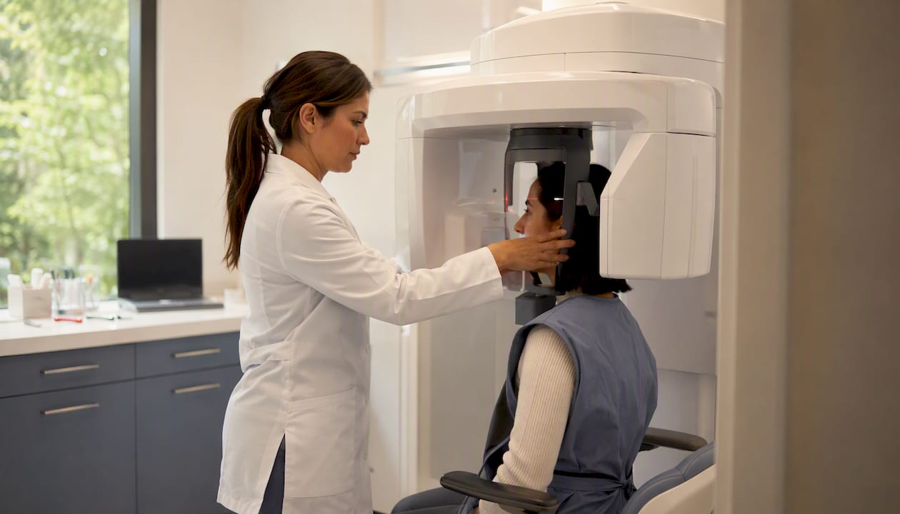
Pro Tip: Ask to see your CBCT images and the 3D treatment plan. Request the specific numbers your surgeon used: bone height, width, and cortical thickness at each proposed implant site. "Your bone looks okay" is not a clinical measurement.
Why do people lose jawbone in the first place?
Jawbone resorption after tooth loss is not a slow, gradual process. The alveolar bone that surrounds tooth roots exists primarily to support those roots. Once a tooth is removed, the bone no longer receives the mechanical stimulation it needs, and resorption begins within weeks. Width loss tends to outpace height loss in the first year, which is why a ridge that looks adequate on a 2D X-ray can be surprisingly narrow on CBCT.
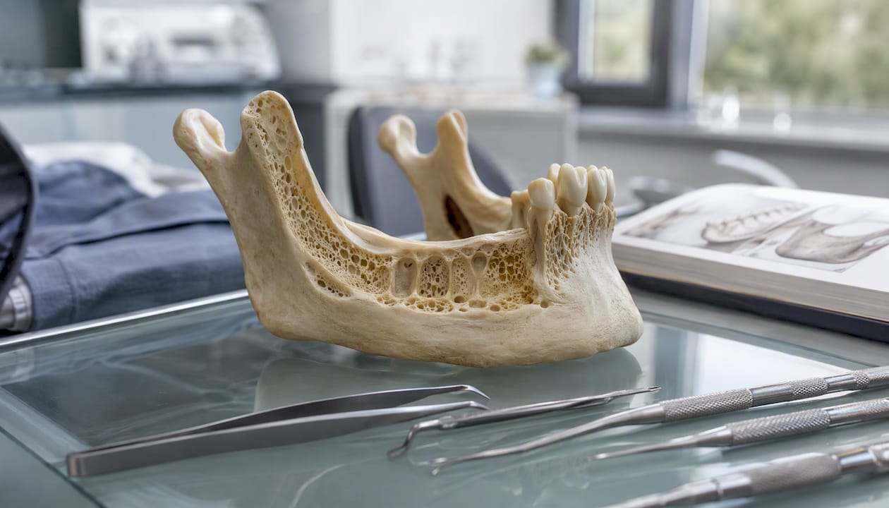
Local causes of bone loss include periodontal disease (which destroys the bone supporting teeth while they are still present), chronic infection or abscess, trauma, and failed implants. Systemic contributors include age-related bone density decline, osteoporosis, and medications that alter bone metabolism. Long-term denture use accelerates this process, which is why many denture wearers arrive at an implant consultation with significantly less bone than they expect.
Some causes are modifiable. Untreated gum disease can be resolved before surgery. Smoking can be stopped. Blood sugar in diabetic patients can be optimized. Others require medical coordination rather than lifestyle change: patients on bisphosphonates or Denosumab need a physician review before bone surgery, and patients who have received radiation to the head and neck require specialist evaluation because radiation damages the blood supply that bone healing depends on.
What are your options when jawbone is insufficient?
Insufficient bone does not automatically disqualify you from implants. It changes the sequence and sometimes the strategy. Here are the main pathways:
- Socket preservation: Placing graft material in the extraction socket immediately after tooth removal to slow resorption and maintain ridge dimensions for future implant placement.
- Particulate bone graft: Granular graft material packed into a defect site to build volume. Commonly used for moderate deficiencies.
- Block graft: A solid piece of bone (typically harvested from the chin or ramus, or sourced as an allograft) used for larger horizontal or vertical deficiencies.
- Ridge augmentation: A broader category covering lateral and vertical procedures to rebuild a resorbed ridge before implant placement.
- Lateral sinus lift (sinus floor elevation): An approach through a window in the lateral sinus wall to lift the sinus membrane and pack graft material below it, creating height in the posterior upper jaw.
- Transcrestal sinus lift: A less invasive approach through the implant osteotomy site, used when only a few millimeters of additional height are needed.
- Zygomatic implants / All-on-4 alternatives: For patients with severe posterior maxillary bone loss, zygomatic implants anchor into the cheekbone (zygoma) rather than the alveolar ridge, bypassing the need for sinus grafting entirely. These are specialist procedures. Patients with severe bone resorption have real options through these approaches.
Graft material comparison
| Graft type | Source | Typical healing window | Common use case |
|---|---|---|---|
| Autograft | Patient's own bone (chin, ramus, hip) | Several months | Large defects; gold standard for volume |
| Allograft | Donor human bone (processed) | Several months | Moderate defects; avoids second surgical site |
| Xenograft (e.g., DBBM) | Bovine or porcine bone mineral | Several months or longer | Sinus lifts, socket preservation |
| Synthetic (alloplast) | Calcium phosphate or similar | Several months | Minor defects; predictable resorption profile |
A randomized clinical trial comparing 5-month versus 8-month healing in staged sinus floor elevation with deproteinized bovine bone mineral found no significant difference in new bone formation between groups, though long-term implant survival effects remain unclear. That variability is worth discussing with your surgeon: some clinicians use shorter healing windows successfully with specific materials, but the objective endpoint (radiographic confirmation of adequate new bone) should be defined before you accept a faster timeline.
Staged vs. simultaneous placement is a decision driven by the amount of primary stability achievable at the time of grafting. When a surgeon can achieve adequate mechanical stability placing the implant at the same time as the graft, simultaneous placement shortens overall treatment time. When the defect is too large for reliable initial stability, a staged approach (graft first, implant after confirmed healing) is the safer path.
How to prepare your jaw and body before implant surgery
Preparation is not just paperwork. The steps you take in the weeks and months before surgery directly affect how well your bone heals and whether osseointegration succeeds.
Medical optimization:
- Disclose all medications, especially bisphosphonates, Denosumab, corticosteroids, anticoagulants, and immunosuppressants. Some require a physician-supervised drug holiday before bone surgery.
- If you have diabetes, work with your physician to bring HbA1c into a controlled range. Uncontrolled blood sugar impairs bone healing and increases infection risk.
- Report any history of head or neck radiation. Radiation-damaged bone heals poorly and may require hyperbaric oxygen therapy as an adjunct.
- Disclose immune status and any recent systemic illness.
Lifestyle steps:
- Stop smoking and all nicotine products as early as possible before surgery. The cellular mechanism matters here: nicotine increases markers tied to osteoclast activity and decreases osteoblast numbers, directly inhibiting osseointegration. The cessation window should be discussed with your surgeon relative to your grafting and loading schedule, not just a generic "quit before surgery" instruction.
- Limit alcohol in the weeks before and after surgery. Alcohol interferes with bone remodeling and increases bleeding risk.
- Weight-bearing exercise supports systemic bone density, though it will not rebuild a resorbed ridge on its own.
Nutrition:
- Adequate calcium, vitamin D, and protein support bone healing. These are baseline requirements, not substitutes for surgical grafting when volume is insufficient.
- Ask your surgeon whether a vitamin D level check before surgery is appropriate. Deficiency is common and correctable.
Dental steps:
- Treat any active gum disease or infection before grafting or implant placement. Bacteria present at a graft site significantly increase failure risk.
- Maintain meticulous oral hygiene in the weeks leading up to surgery to reduce the bacterial load around proposed surgical sites.
Pro Tip: Bring a printed medication list to your consultation, including over-the-counter supplements. Fish oil, vitamin E, and aspirin all affect bleeding. Your surgeon needs the full picture.
Who is not a candidate, and what happens then?
Most people with insufficient bone are not disqualified from implants. They are paused for staged treatment. The distinction matters.
High-risk factors that typically require medical clearance or treatment modification:
- Uncontrolled diabetes (elevated HbA1c)
- Active periodontal disease or untreated oral infection
- Recent head and neck radiation (typically within the past year, though the threshold varies by dose and field)
- History of IV bisphosphonate therapy (used for cancer treatment, not the oral doses prescribed for osteoporosis, which carry lower risk)
- Heavy active smoking without a willingness to cease
- Severe immunosuppression
Relative contraindications (factors that increase risk but do not automatically prevent treatment) include controlled systemic disease, oral bisphosphonate use, and moderate smoking history. These require honest discussion about risk, not automatic disqualification.
When clinicians recommend staged treatment, the typical sequence is: resolve the systemic or local risk factor, complete grafting, confirm healing radiographically, then place implants. That sequence can add 6–18 months to the overall timeline. The trade-off is a substantially higher probability of long-term success versus attempting implant placement in compromised conditions and facing removal and reattempt later.
What are the realistic timelines and costs for bone procedures?
Bone procedures add time and cost to implant treatment. Here is a realistic reference table:
| Procedure | Typical healing window | Notes |
|---|---|---|
| Socket preservation | Several months | Performed at extraction; maintains ridge for future implant |
| Particulate bone graft | Several months | Staged placement after confirmed healing |
| Block graft | Several months | Larger defects; requires second surgical site or allograft |
| Lateral sinus lift | Several months or longer | Posterior upper jaw; healing confirmed radiographically |
| Implant osseointegration | Several months | After placement; varies by bone quality and loading protocol |
| Immediate loading (same-day teeth) | Provisional teeth day of surgery; final restoration after osseointegration | Requires adequate primary stability at placement |
Cost context: Full-arch implant treatment at Forever Smiles Implant Center starts from $19,000 per arch. That price includes planning, surgery, same-day provisional teeth, final custom zirconia restorations, and follow-up care. Bone grafting procedures, when required, are factored into the individualized treatment plan. See the full-arch implant cost and financing page for details.
Cost drivers beyond the base procedure include: CBCT imaging and 3D planning, surgical specialist fees, anesthesia (a board-certified MD anesthesiologist adds safety for complex cases), graft material type and volume, in-house lab fabrication of zirconia restorations, and geographic market factors. Getting a written, itemized estimate before committing to treatment is not optional. Any provider unwilling to put the plan and price in writing is a red flag.
Practical steps to preserve or rebuild bone before implants
The most effective thing you can do after losing a tooth is act quickly. Socket preservation performed at the time of extraction is far simpler and less expensive than rebuilding a resorbed ridge later. Replacing missing roots promptly, whether with implants or temporary measures that maintain space, slows the resorption cascade.
Beyond timing, the modifiable factors that most directly affect bone healing potential are:
- Nicotine cessation: The biological case is clear. Research shows nicotine inhibits osseointegration by altering osteoblast and osteoclast activity, with statistically significant differences compared to non-smokers. Cessation timing relative to grafting and loading is a nuanced conversation, not a single universal rule. Your surgeon should give you a specific window, not just a general recommendation.
- Blood sugar control: Optimizing HbA1c before surgery improves bone healing outcomes in diabetic patients.
- Medication review: Work with your prescribing physician before stopping or modifying any bone-modifying medication. Never discontinue bisphosphonates without physician guidance.
- Oral hygiene: Keeping the graft site clean is the single most controllable factor in graft survival post-operatively.
Supplements alone will not rebuild a resorbed ridge. Calcium and vitamin D support the healing process but cannot create bone volume where none exists. Surgery does that.
Questions to bring to your implant consultation
Walk in with these questions written down. A surgeon who cannot answer them specifically is not planning your case with enough precision.
- Do I need a CBCT scan, and will you show me the measurements from it?
- What are the specific bone height, width, and cortical thickness at each proposed implant site?
- Am I a candidate for immediate implant placement, or do I need grafting first?
- If grafting is recommended, what material will you use and what is the healing timeline before implant placement?
- What is the loading schedule: immediate provisional teeth, delayed loading, or staged?
- How do my current medications affect the surgical plan or timeline?
- What is the specific cessation window you recommend for nicotine given my grafting schedule?
- What happens if the graft does not integrate as expected?
- What are the measurements and objective criteria you will use to confirm I am ready for implant placement after grafting?
- Can I have a written treatment plan with the timeline and itemized cost estimate?
Pro Tip: Request a printed copy of your CBCT-based 3D plan showing the proposed implant positions and measurements. This is your record of the clinical rationale for the treatment plan.
What a specialist implant pathway looks like for minimal-bone patients
A realistic care pathway for a patient presenting with significant bone loss at a specialist center follows a predictable sequence, though the specifics vary by case.
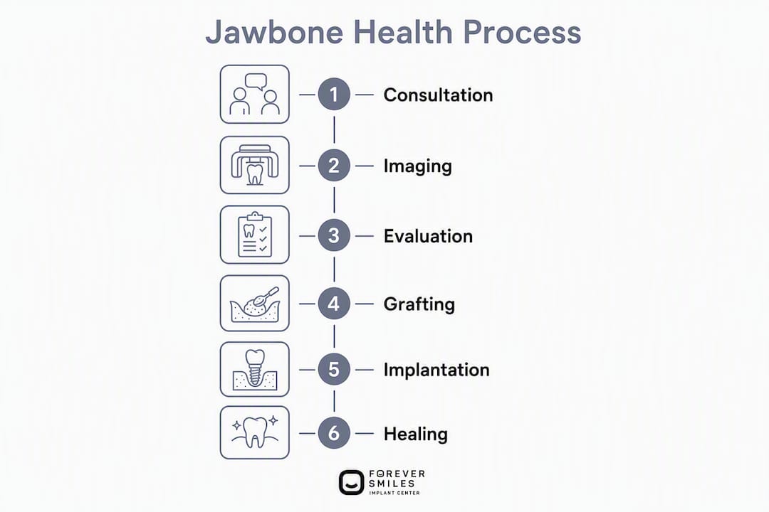
Stage 1: Consultation and CBCT imaging. The surgeon reviews medical history, performs a clinical exam, and acquires a CBCT scan. Bone height, width, cortical thickness, and proximity to anatomical landmarks are measured at each proposed implant site. A 3D treatment plan is generated.
Stage 2: Grafting (if indicated). Depending on the defect, this may be socket preservation, a particulate or block graft, a sinus lift, or a combination. The graft material is selected based on defect size, patient health, and surgeon preference. Healing is confirmed radiographically before proceeding.
Stage 3: Implant placement. Once adequate bone volume is confirmed, implants are placed according to the 3D plan. For full-arch cases with sufficient primary stability, provisional teeth may be delivered the same day.
Stage 4: Osseointegration and final restoration. After the osseointegration period (typically 3–6 months), final custom zirconia restorations are fabricated in the in-house lab and delivered.
Dr. Brian Young is a residency-trained surgical specialist with more than 20 years of experience and over 28,000 implants placed. He performs full-arch implant surgery daily, trains surgeons nationally, lectures on full-arch technique, and sits on the editorial board of Implant Practice US. The clinic operates with a board-certified MD anesthesiologist on staff and an in-house dental lab, meaning every stage of the process from 3D imaging and planning through final zirconia restorations happens under one roof with one accountable team.
Key Takeaways
Sufficient jawbone volume and cortical thickness are prerequisites for osseointegration. When bone is lacking, grafting or alternative implant strategies make treatment possible but add months to the timeline.
| Point | Details |
|---|---|
| CBCT is the standard | 3D imaging measures bone height, width, cortical thickness, and proximity to nerves and sinuses before any surgical plan is finalized. |
| Cortical thickness matters | Research shows cortical thickness significantly influences primary implant stability, not just overall bone density. |
| Grafts take months | Transplanted bone typically requires several months to grow enough new bone to support implants; staged protocols are common. |
| Nicotine inhibits healing | Nicotine decreases osteoblast numbers and increases osteoclast activity, directly interfering with osseointegration. |
| Forever Smiles Implant Center | Specialist center in Jacksonville offering CBCT-based planning, staged grafting, zygomatic implants, and full-arch reconstruction from $19,000 per arch. |
What surgeons actually prioritize when bone is low
The conversation most patients expect at an implant consultation is "do you have enough bone?" The conversation that actually matters is more specific than that.
When a patient presents with minimal bone, the first priority is not deciding whether to proceed. It is understanding why the bone is gone and whether the conditions that caused the loss are still active. A patient with uncontrolled periodontal disease who gets a graft without treating the disease first is setting up that graft to fail. A patient on IV bisphosphonates who has not had a physician review is a surgical risk that no amount of imaging can mitigate.
The second priority is mechanical: what primary stability is achievable at the proposed sites, and does that stability support immediate loading or require a staged approach? This is where cortical thickness becomes the operative variable. A jaw with low overall density but intact cortical walls can still provide the mechanical grip needed for initial osseointegration. A jaw with good density but thin cortical bone at the crest may not. That distinction drives the loading schedule, which in turn drives the entire treatment timeline.
What patients underestimate is how much the sequence of decisions matters. Grafting before implants is not a detour. It is the plan. Patients who understand that tend to have better outcomes, not because the biology is different, but because they follow the protocol rather than pushing for shortcuts that compromise the result.
The cases I find most rewarding are the ones where a patient was told elsewhere they were not a candidate. Often, they were told that because the provider lacked the tools or experience to plan around the deficiency. With CBCT-based planning, zygomatic implant options, and a staged grafting protocol, the answer is rarely "no." It is usually "here is the sequence."
Forever Smiles Implant Center: built for the cases other providers decline
Patients who arrive at Forever Smiles Implant Center with severe bone loss, previously failed implants, or a history of being told they are not candidates are not edge cases here. They are the core of what the practice does every day.
The workflow is built around the complexity: CBCT-based 3D planning at consultation, staged grafting protocols when needed, zygomatic and pterygoid implant options for patients with no posterior maxillary bone, and full-arch reconstruction delivered under one roof with a board-certified MD anesthesiologist and an in-house zirconia lab. Full-arch treatment starts from $19,000 per arch and includes planning, surgery, same-day provisional teeth, final restorations, and follow-up care.
If you have been told your bone is insufficient, or if you are simply ready to know exactly where you stand, the right next step is a CBCT consultation. Schedule your 3D imaging consultation at Forever Smiles Implant Center and leave with a written plan, documented measurements, and a clear timeline.
Useful sources and further reading
The claims in this guide draw from peer-reviewed research, systematic reviews, and clinical guidelines. The sources below are the primary references:
- Efficacy of clinical methods to assess jawbone tissue prior to and during endosseous dental implant placement: systematic literature review (NCBI Bookshelf / DARE)
- Retrospective analysis of dental implant stability in relation to mandibular bone density and cortical thickness (PMC)
- Randomized clinical trial on sinus augmentation using DBBM: 5-month vs. 8-month healing (PubMed)
- Dental implant surgery overview (Mayo Clinic)
- Implant Consultation and 3D Imaging, Jacksonville, FL (Forever Smiles Implant Center)
- Full-Mouth Dental Implants, Jacksonville, FL (Forever Smiles Implant Center)
Bring printed copies of any existing X-rays, CBCT scans, or medical records to your consultation. The more clinical data your surgeon has at the first appointment, the more specific and accurate the treatment plan will be.
This article provides general educational information about jawbone health and dental implant surgery. It is not a substitute for professional medical or dental advice. Confirm your specific situation and treatment options with a qualified surgical specialist.